CVI
A program of Crown Valley Imaging · Newport Beach

Private Executive Portal · Newport Beach
Executive Full-Body MRI Screening in Newport Beach
Radiologist-led preventive imaging for Orange County executives who want early structural signal and rapid, physician-ready clarity.
- See risk sooner. Act earlier.
- Executive-grade preventive imaging.
- AI-assisted MRI protocols for speed and image consistency.
- Your annual health baseline, upgraded.
40-90 min
Scan window
Tier-dependent MRI depth
3 tiers
Program architecture
Signature, Elite, Reserve
30-day
Scheduling window
Concierge logistics for any follow-up your physician orders
Private
Concierge communication
Discretion-first workflow

Premium preventive imaging
Three program tiers built around MRI precision, optional CT context, and continuity planning.
Pricing at a glance · Click any tier for full inclusions
Clinical Standards
Built on institutional-grade imaging foundations
Named Radiology Leadership
Every scan is read by a named CVI radiologist
Four subspecialty fellowship-trained physicians. Seventy-plus years of combined practice experience. Every Peak Prevention scan is read in-house by one of our named radiologists.
Talal Beydoun, MD
Medical Director · Founder
Neuroradiology · Interventional Radiology
Dr. Beydoun founded Crown Valley Imaging in 2002 and brings more than five decades of clinical, academic, and leadership experience in radiology. He holds American Board of Radiology Certificates of Added Qualification (CAQ) in both Neuroradiology and Interventional Radiology, and formerly served as Clinical Professor of Radiological Sciences at UC Irvine Medical Center, where he was named Teacher of the Year (2000–2001). He also actively reads musculoskeletal cases. At CVI he leads clinical quality, imaging protocols, physician education, and the Peak Prevention Program's dual-specialist read model.
- Board-Certified, American Board of Radiology (1974)
- ABR CAQ in Neuroradiology (1999)
- ABR CAQ in Interventional Radiology (2001)
- Residency & Fellowship: UT MD Anderson Hospital & Tumor Institute
- Former Clinical Professor, UC Irvine Medical Center (1998–2002)
- Teacher of the Year, UCI Fellows & Residents (2000–2001)
- Active MSK reads
- Five decades of radiology practice · Founder, Crown Valley Imaging (2002)

Alireza Namini, MD
Neuroradiologist
Neuroradiology
Dr. Namini completed his Neuroradiology fellowship at the Keck School of Medicine of USC after a diagnostic radiology residency at Hahnemann University Hospital in Philadelphia — a Level I trauma center with high-volume emergency imaging exposure. He earned his MD from Drexel University College of Medicine. At CVI he leads brain, spine, and head-and-neck interpretation for the Peak Prevention Program, including high-priority findings such as aneurysms, white-matter change, and cervical stenosis, and brings experience in minor interventional and spinal pain management procedures.
- Board-Certified Radiologist (ABR)
- Fellowship: Neuroradiology, Keck School of Medicine of USC
- Residency: Diagnostic Radiology, Hahnemann University Hospital, Philadelphia (Level I trauma center)
- MD, Drexel University College of Medicine
- BS, Biology (Magna Cum Laude), California State University Fullerton
- Subspecialty focus: brain, spine, head & neck

James R. Fernandez, MD
Neuroradiologist
Neuroradiology · Imaging Informatics
Dr. Fernandez completed his Neuroradiology fellowship at UT Southwestern Medical Center after a Diagnostic Radiology residency at LAC+USC Medical Center, with additional mini-fellowships in Breast and Thoracic imaging. Before clinical training, he was an NIH NIBIB T32 Training Fellow in Imaging Informatics at the USC Image Processing and Informatics Laboratory (IPILab), where his research focused on computer-aided detection of intracranial hemorrhage and quantitative imaging analysis. He earned his MD from the John A. Burns School of Medicine at the University of Hawaii and his BS in Biomedical Engineering from USC. At CVI he supports neuroradiology interpretation for the Peak Prevention Program — brain, spine, and head-and-neck reads — and contributes to the integration of AI-assisted detection methodologies into the read workflow.
- Board-Certified Radiologist (ABR)
- ABR CAQ in Neuroradiology
- Fellowship: Neuroradiology, UT Southwestern Medical Center
- Residency: Diagnostic Radiology, LAC+USC Medical Center
- Mini-Fellowships: Breast Imaging & Thoracic Imaging, LAC+USC
- NIH NIBIB T32 Training Fellow, USC Image Processing & Informatics Laboratory
- MD, John A. Burns School of Medicine (University of Hawaii)
- BS, Biomedical Engineering, University of Southern California
- Published in SPIE Medical Imaging proceedings (computer-aided detection / informatics)

Tan Nguyen, MD
Musculoskeletal & General Radiologist
Musculoskeletal Radiology
Dr. Nguyen completed his Musculoskeletal Imaging fellowship at UC San Diego following a Diagnostic Radiology residency at UC Irvine and an MD from the David Geffen School of Medicine at UCLA. His training included full-modality MSK interpretation across MRI, CT, and radiographs with daily exposure to arthrograms and image-guided procedures. A former NIH Clinical Research Training Program fellow with peer-reviewed publications in Radiology, the British Journal of Radiology, and BMC Musculoskeletal Disorders, he leads joint, soft-tissue, and body-composition interpretation for the Peak Prevention Program, including DEXA analysis in the Reserve tier.
- Board-Certified Radiologist (ABR)
- Fellowship: Musculoskeletal Imaging, UC San Diego
- Residency: Diagnostic Radiology, UC Irvine
- MD, David Geffen School of Medicine at UCLA
- NIH Clinical Research Training Program Research Fellow
- Published in Radiology, Br J Radiol, BMC Musculoskeletal Disorders
All CVI radiologists are board-certified by the American Board of Radiology with fellowship training in their respective subspecialties.
Elite and Reserve tiers are read by the subspecialist whose domain best matches the clinical context. Reserve includes a dual-specialist read model — with Neuroradiology, Interventional Radiology, and MSK expertise on the CVI bench, brain and spine findings and musculoskeletal or body-composition signal are each reviewed by a physician fellowship-trained in the relevant discipline.
American College of Radiology Accreditation
ACR Accredited across both MRI and CT
CVI's facility holds American College of Radiology accreditation for both Magnetic Resonance Imaging and Computed Tomography — a peer-reviewed verification of equipment, safety, personnel qualifications, and image quality standards. ACR accreditation is the recognized benchmark for clinical imaging quality in the United States.
MRI Accredited Facility
ACR Accredited — Magnetic Resonance Imaging
Peer-reviewed verification of equipment, safety, personnel qualifications, and image quality.
CT Accredited Facility
ACR Accredited — Computed Tomography
Peer-reviewed verification of equipment, safety, personnel qualifications, and image quality.
ACR accreditation does not constitute a diagnostic or outcomes guarantee. It verifies that the facility meets published quality standards at the time of review.
Program Tiers
Three preventive imaging programs, one radiology team
Three focused pathways: Signature, Elite, and Reserve.
Signature
$040-minute whole-body MRI (brain included)
Annual baseline MRI with brain imaging. The preventive core for a minimal time burden.
Best for: Leaders who want a high-quality annual baseline with minimal time burden.
Read model: Single subspecialty radiologist interpretation
Not Included
- Head-to-pelvis MRI including brain screening
- Structured specialist interpretation
- Prioritized findings report routed to your referring physician
- Concierge scheduling for any follow-up your physician orders
Upgrade signal: Upgrade to Elite for deeper neuro/spine coverage and a radiologist results walkthrough.
Elite
$060-minute extended MRI
Extended MRI depth with a 30-day concierge scheduling window after your physician receives the report.
Best for: Executives who want broader MRI depth, especially neuro and spine context.
Read model: Enhanced subspecialty interpretation with a radiologist results walkthrough — findings routed to your referring physician for clinical decisions
Not Included
- Advanced sequences
- Radiologist results walkthrough
- 30-day concierge scheduling window for physician-ordered follow-up
- Executive summary report routed to your referring physician
Upgrade signal: Upgrade to Reserve for MRI+CT+CAC+DEXA and dual-specialist interpretation.
Reserve
$0Two-day comprehensive program · MRI + MRA/MRV + CT + CAC + DEXA
A choreographed two-day program adding comprehensive MRA/MRV vascular imaging to the whole-body MRI, then pairing it with CT, coronary calcium, and DEXA at our Mission Viejo facility. Executive private transportation between visits is included.
Best for: Principals and family-office operators who want maximum signal and a fully orchestrated two-day experience.
Read model: Dual-specialist read model: Neuroradiologist + MSK radiologist
- Everything in Elite
- Day 1 (~2.5 hours): 60-minute whole-body 3T MRI + 90-minute comprehensive MRA/MRV at CVI Newport Beach (4501 Birch St)
- Comprehensive MRA: Circle of Willis, carotid arteries, thoracic aorta, abdominal vasculature
- MRV: cerebral venous sinuses and major venous return
- Day 2: Full-body low-dose CT (thorax/abdomen/pelvis), Coronary Calcium Score, and DEXA at CVI Mission Viejo
- Executive private transportation to and between both visits
Imaging Environment
MRI-first suite design

Diagnostic Signal
CT + MRI signal layering
What Is Included / Not Included
Tier-level inclusion matrix
Transparent scope view for quick planning.
Signature
Elite
Reserve
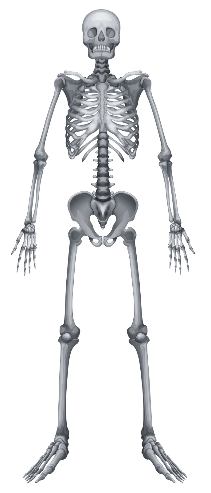
Anatomical Coverage · Visualized
What each tier actually scans
Toggle between tiers. Reserve activates a live vascular overlay showing the MRA / MRV pathways.
Elite Tier
Extended MRI depth with a 30-day concierge scheduling window after your physician receives the report.
Brain + Head/Neck
Neurostructural screening and head/neck soft-tissue signal review.
Spine
Degenerative and structural pattern review across major spinal segments.
Thorax + Cardiovascular Context
Chest structural signal with cardiovascular risk-context layers.
Abdomen
Organ-level structural review for major abdominal systems.
Pelvis
Pelvic-organ and deep soft-tissue structural mapping.
Musculoskeletal + Composition
Vascular — MRA + MRV
Illustrative anatomical reference only. Exact sequences, coverage boundaries, and interpretation methodology are protocol-specific and adjusted by the reading radiologist based on clinical context.
Structure-Level Detail
Every structure reviewed, by tier
A granular view of the specific anatomical structures covered in each tier's protocol.
| Region / Structure | Signature | Elite | Reserve |
|---|---|---|---|
Brain + Head/NeckNeurostructural screening and head/neck soft-tissue signal review. | |||
| Brain parenchymal structure and ventricular patterning | |||
| Pituitary/sellar region overview | |||
| Sinus and major head/neck soft-tissue context | |||
| Thyroid-region incidental structural signal | |||
SpineDegenerative and structural pattern review across major spinal segments. | |||
| Cervical disc and canal-level signal | |||
| Thoracic vertebral alignment overview | |||
| Lumbar degeneration and stenosis patterning | |||
| Paraspinal soft-tissue context | |||
Thorax + Cardiovascular ContextChest structural signal with cardiovascular risk-context layers. | |||
| Aortic caliber and thoracic vascular morphology | |||
| Mediastinal and pulmonary structural overview | |||
| CT Coronary Calcium Score (heart plaque burden): included in Reserve, available as add-on in Signature/Elite | |||
| Cardiometabolic structural patterning | |||
AbdomenOrgan-level structural review for major abdominal systems. | |||
| Liver, pancreas, spleen, and adrenal architecture | |||
| Renal morphology and incidental lesion context | |||
| Gallbladder and biliary-tract structural signal | |||
| Major abdominal soft-tissue findings prioritization | |||
PelvisPelvic-organ and deep soft-tissue structural mapping. | |||
| Bladder and pelvic soft-tissue architecture | |||
| Reproductive-organ structural context | |||
| Pelvic vascular/lymphatic pattern indicators | |||
| Incidental findings flagged and prioritized in the report to your referring physician | |||
Musculoskeletal + CompositionBone, joint, and composition-related structural signal layers. | |||
| Major joint and periarticular soft-tissue patterning | |||
| DEXA body-fat percentage and lean-mass profile: included in Reserve, available as add-on in Signature/Elite | |||
| Skeletal asymmetry and loading trend indicators | |||
| Priority mapping in the report to help your physician plan next steps | |||
Vascular — MRA + MRVNon-contrast arterial and venous imaging for early vascular-risk visibility. | |||
| 3D TOF MRA — Circle of Willis (intracranial arteries, aneurysm context) | |||
| MRA — carotid and vertebral arteries (stenosis, dissection screen) | |||
| MRV — cerebral venous sinuses (venous return patterning) | |||
| Non-contrast MRA — thoracic aorta (aneurysm / dissection screen) | |||
| Non-contrast MRA — abdominal aorta + renal arteries | |||
Dual-Modality Intelligence
MRI + CT for complementary structural signal
MRI and CT contribute different but complementary forms of structural intelligence, improving risk visibility in complex decision environments.
MRI Strength
Soft-tissue, neurologic, and organ-level signal without ionizing exposure.
CT Strength
Fast structural and calcification visibility for thoracic and vascular context.
Integrated View
Combined interpretation sharpens triage and prioritization pathways.
Add Coronary Calcium or DEXA
Add CAC or DEXA to your imaging program
Member pricing — available only when bundled with a Peak Prevention tier.
Select base tier
Add-ons
Bundle rule
When CAC and DEXA are selected together, bundled pricing applies. $299.
Member pricing — these CAC and DEXA rates are part of the Peak Prevention program. Standalone à la carte CAC and DEXA at Crown Valley Imaging are priced separately.
Estimated Total
Signature$1,999
Add-ons selected$0
Tier + CAC + DEXA benchmark
Signature + CAC + DEXA$2,298
Elite + CAC + DEXA$3,598
Reserve (CAC + DEXA included)$9,999
HSA / FSA Pathways Supported
Many CVI patients use HSA or FSA funds to pay for their scan. Eligibility is determined by your plan administrator and IRS rules. We provide an itemized receipt and, when clinically appropriate, can coordinate a Letter of Medical Necessity through a licensed provider. Interest-free payment plans are available.
HSA and FSA eligibility is determined by your plan administrator under IRS rules and is not guaranteed. CVI provides an itemized receipt and, when clinically appropriate, can coordinate a Letter of Medical Necessity through a licensed provider. Reimbursement is not guaranteed. Payment plans are subject to approval and may involve third-party financing with separate terms. Prices shown are for the CVI preventive program only and do not include downstream diagnostic studies, specialist consultations, or treatment that may be indicated after your scan.
Clinical Boundaries & Safety
What this program is, and what it is not
Transparency supports better decisions. This program is a preventive intelligence layer that complements physician-led care.
This Program Is
- An elective preventive imaging program focused on early structural visibility
- A radiologist-interpreted, physician-routed reporting model — every report goes to your referring physician, who owns all clinical decisions and follow-up care
- Concierge scheduling and logistical coordination for any imaging your physician orders at our facility
- Open only to participants who designate a referring or ordering physician of record (primary care, internist, or specialist)
This Program Is Not
- Not diagnostic medicine, emergency care, or urgent care
- Not a replacement for routine guideline-based screening (colonoscopy, mammography, Pap, PSA, skin, dental, vision) directed by your physician
- Not a substitute for direct diagnosis, treatment, or follow-up care by your physician — those decisions remain with them
- Not a guarantee of disease detection, disease prevention, or risk reduction
- Not the establishment of a physician-patient relationship between you and CVI
Contraindication Categories
- MRI contraindications may apply for certain implanted devices and ferromagnetic hardware
- Pregnancy and other clinical factors require physician discussion before enrollment
- Severe claustrophobia may require preparatory planning
- Patients with acute symptoms should seek emergency or urgent care — not preventive imaging
Operational Timelines
Priority findings
Communicated to your referring physician same-day when clinically indicated
Structured report release
Routed to your referring physician within 48 business hours or less
Concierge scheduling window
30 days of logistical support for any follow-up imaging your physician orders (applicable tiers)
This is an elective preventive imaging service and is not diagnostic medicine. CVI radiologists interpret the study and route the report to your designated referring or ordering physician; participation does not create a physician-patient relationship with CVI. Imaging has inherent limitations, including false-positive and false-negative findings. No guarantee of disease detection, prevention, or outcome is made. All clinical decisions are made by your licensed physician. If you are experiencing acute symptoms, call 911 or go to the nearest emergency department.
FAQ
Executive preventive imaging questions
Clear answers for intake, scope, and follow-through.
This is an elective preventive imaging program focused on early structural visibility. It is not diagnostic medicine, emergency care, or a substitute for routine medical care. Participation does not establish a physician-patient relationship. All diagnosis and treatment decisions remain with your licensed physician team.
Ready when you are
Start a conversation with our team.
We'll align tier, scheduling, and follow-through in one short intake. No medical detail required up front.
13+
Organs Screened
Head-to-pelvis MRI coverage across all major anatomical systems
3T
MRI Grade
Research-grade magnetic field strength for superior tissue contrast
<60 min
Scan Duration
Efficient protocols designed for compressed executive schedules
48 hr
Report Turnaround
Physician-ready structured reports delivered within two business days
For many cancers and vascular conditions, published stage-specific survival data show meaningfully better outcomes when disease is identified at earlier stages. Preventive imaging is one of several tools physicians use toward that goal; it is not a guarantee of detection or outcome.